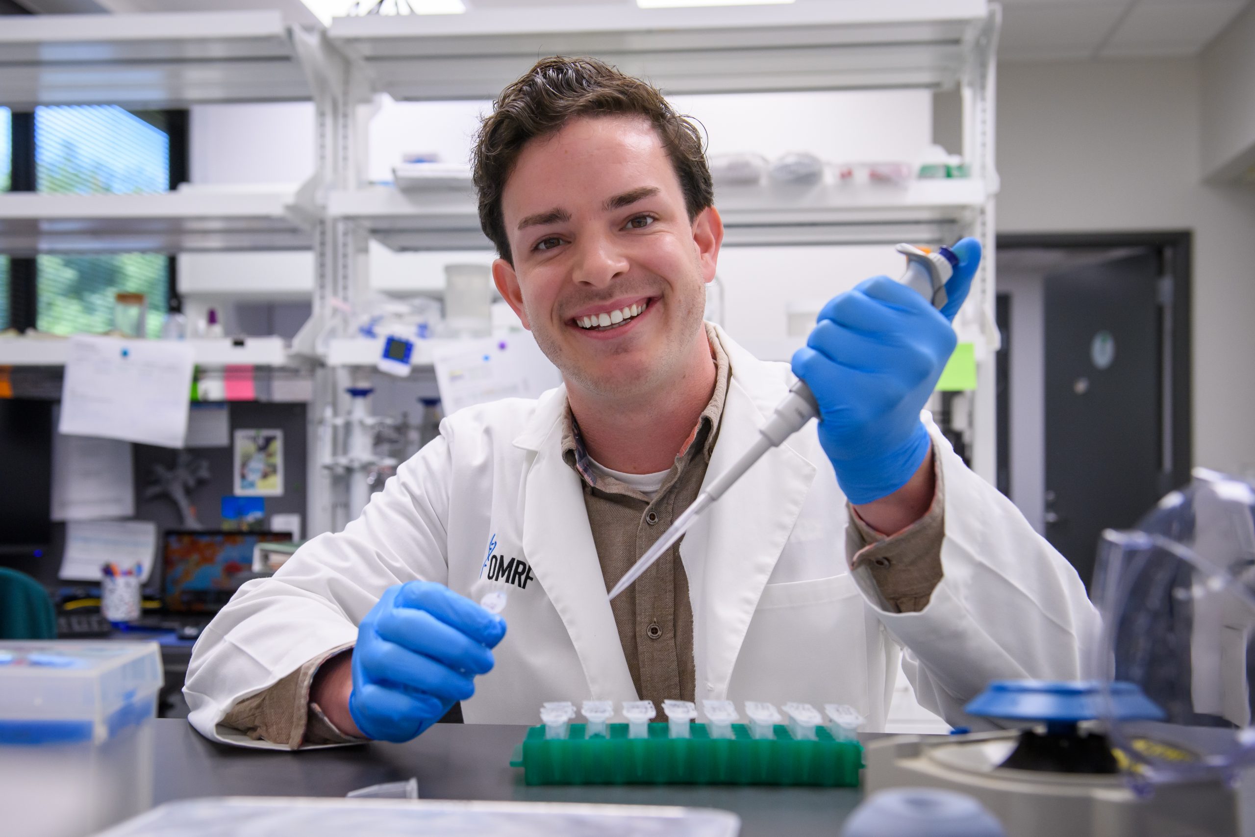
Zachary Hettinger, Ph.D.
Assistant Professor
Aging & Metabolism Research Program
My 101
The guiding framework of our research is that healthy muscles lead to healthy aging. Skeletal muscle is the “motor” for movement, but during aging often becomes dysfunctional, leading to mobility limitations and lower life satisfaction. In our lab, we tap into the basic biology of aging and muscle to discover new ways to help older adults keep moving to maintain the independent lifestyle they deserve.
Specifically, our lab researches how aging and a sedentary lifestyle promotes the accumulation of fat and connective tissue inside of skeletal muscle, and how their accumulation impairs the muscle’s ability to produce force. Of particular interest are how stem and progenitor cells and their dysfunction contribute to these issues, and whether targeting these cell populations therapeutically can rescue muscle function. Ultimately, it is our goal that discoveries made in our lab translate to tangible treatment options that promote muscle health during aging.
Research
Muscle contraction is carried out by contracting myofibers ensheathed in an extracellular matrix (ECM) aiding in force production. Muscle-resident stromal cells termed fibro-adipogenic progenitor cells (FAPs) are responsible for maintaining the ECM, endowing it with the material properties conducive to force transmission. With aging, however, FAPs lose their homeostastic roles and contribute to the accumulation of their fibrogenic and adipogenic progeny. The consequence of FAP dysfunction is a transformed cellular neighborhood that no longer serves to support muscle function.
Our lab examines muscle-resident FAP dysfunction with aging through both a systems-based and cellular lens.
From a systems perspective, our lab focuses on how changes in circulating endocrine signals with age influence muscle-resident FAPs to adopt fibrogenic and adipogenic lineages. The gradual loss of androgens in males and the more rapid loss of estrogens in females during aging leads to breakdowns in endocrine feedback regulatory loops, which dramatically alters the circulating hormonal milieu. Our lab examines how the transition from a “youthful” to an “aged” circulating hormone profile influences the ability of FAPs to support muscle homeostasis using a combination of animal models and cell culture assays.
From a cellular perspective, we also research whether aging impairs FAPs ability to maintain a “youthful” gene regulatory network (GRN) by altering the intracellular biophysical environment. The maintenance of a functional GRN relies of the efficient processing of cell state-specific genetic information, which, at its core, hinges on the ability of the “right” proteins being able to interact with the “right” genes. Less than 20 years ago, it was discovered that cells are capable of promoting certain macromolecular interactions over others through a process called liquid-liquid phase separation (LLPS). In our lab, we combine single cell genomics with super-resolution microscopy to investigate whether aging impairs the ability of FAPs to undergo LLPS and protect GRN-related genetic material.
Brief CV
Education
B.S., Purdue University, 2016
M.S., Purdue University, 2018
Ph.D., University of Kentucky, 2021
Postdoctoral training, University of Pittsburgh, 2021-2022
Postdoctoral training, Harvard Medical School, 2022-2024
Honors and Awards
Outstanding Musculoskeletal Research Award, Spaulding Rehabilitation Hospital, 2024
Poster Presentation Award, Harvard Medical School, 2023
Research Recognition Award, Cell and Molecular Physiology Section, American Physiology Society, 2023
Travel Award, Oklahoma Geroscience Symposium, 2022
NIH NRSA F31 Predoctoral Fellowship, 2021
Memberships
American Aging Association
Orthopaedic Research Society
American Physiological Society
Alliance for Regenerative Rehabilitation Research and Training
Joined OMRF Scientific Staff in 2024
Publications
Recent Publications
Hughes DC, Hettinger ZR, Deane CS. Androgen-Mediated Regulation of Skeletal Muscle Mass: A Ticking Clock. Function (Oxf), 2025 August, PMID: 40815271, PMCID: PMC12448486
Sklivas AB, Hettinger ZR, Rose S, Mantuano A, Confides AL, Rigsby S, Peelor FF 3rd, Miller BF, Butterfield TA, Dupont-Versteegden EE. Responses of skeletal muscle to mechanical stimuli in female rats following and during muscle disuse atrophy. J Appl Physiol (1985), 2025 January, PMID: 39884317, PMCID: PMC12184423
Gilmer G, Hettinger ZR, Tuakli-Wosornu Y, Skidmore E, Silver JK, Thurston RC, Lowe DA, Ambrosio F. Female aging: when translational models don't translate. Nat Aging. 2023 Dec;3(12):1500-1508. doi: 10.1038/s43587-023-00509-8. Epub 2023 Dec 5. Erratum in: Nat Aging. 2024 Jan;4(1):164. doi: 10.1038/s43587-023-00561-4. PMID: 38052933; PMCID: PMC11099540.
Hettinger ZR, Wen Y, Peck BD, Hamagata K, Confides AL, Van Pelt DW, Harrison DA, Miller BF, Butterfield TA, Dupont-Versteegden EE. Mechanotherapy Reprograms Aged Muscle Stromal Cells to Remodel the Extracellular Matrix during Recovery from Disuse. Function (Oxf). 2022 Mar 24;3(3):zqac015. doi: 10.1093/function/zqac015. PMID: 35434632; PMCID: PMC9009398.
Hettinger ZR, Kargl CK, Shannahan JH, Kuang S, Gavin TP. Extracellular vesicles released from stress-induced prematurely senescent myoblasts impair endothelial function and proliferation. Exp Physiol. 2021 Oct;106(10):2083-2095. doi: 10.1113/EP089423. Epub 2021 Aug 26. PMID: 34333817.
Van Pelt DW, Hettinger ZR, Vanderklish PW. RNA-binding proteins: The next step in translating skeletal muscle adaptations? J Appl Physiol (1985). 2019 Aug 1;127(2):654-660. doi: 10.1152/japplphysiol.00076.2019. Epub 2019 May 23. PMID: 31120811.
Dungan CM, Murach KA, Zdunek CJ, Tang ZJ, Nolt GL, Brightwell CR, Hettinger ZR, Englund DA, Liu Z, Fry CS, Filareto A, Franti M, Peterson CA. Deletion of SA β-Gal+ cells using senolytics improves muscle regeneration in old mice. Aging Cell. 2022 Jan;21(1):e13528. doi: 10.1111/acel.13528. Epub 2021 Dec 13. PMID: 34904366; PMCID: PMC8761017.
Ismaeel A, Van Pelt DW, Hettinger ZR, Fu X, Richards CI, Butterfield TA, Petrocelli JJ, Vechetti IJ, Confides AL, Drummond MJ, Dupont-Versteegden EE. Extracellular vesicle distribution and localization in skeletal muscle at rest and following disuse atrophy. Skelet Muscle. 2023 Mar 10;13(1):6. doi: 10.1186/s13395-023-00315-1. PMID: 36895061; PMCID: PMC9999658.
Contact
Aging and Metabolism Research Program, Mail Stop 46
825 NE 13th Street
Oklahoma City, Oklahoma 73104
Phone: 405-271-1957
E-mail: Zach-Hettinger@omrf.org
For media inquiries, please contact OMRF’s Office of Public Affairs at news@omrf.org.
Lab Staff
Joanna Papinska, Ph.D.
Postdoctoral Scientist
Xiaoyu Ren, Ph.D.
Research Technician IV
Tanisha Goyal
Research Technician II
Heather Spencer
Administrative Assistant III


