Using 3D Models to Tackle Respiratory Illness
Respiratory syncytial virus is the leading cause of pneumonia worldwide. It takes a particularly heavy toll on children, infecting more than half in the first year of life and nearly 100 percent by age 2.
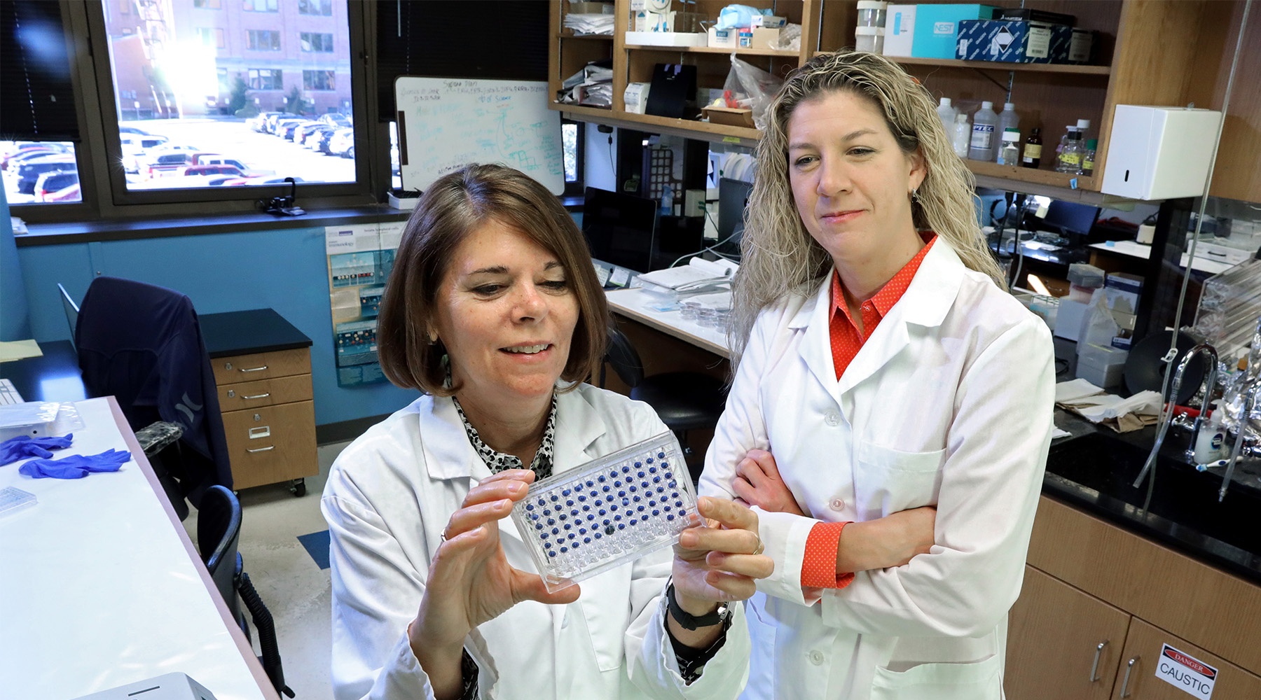
“RSV causes more frequent and more severe infections in infants, compared to adults, and it can be very serious in children, even requiring hospitalization in some cases,” says Dr. Susan Kovats, who studies the virus in her lab at OMRF.
“It’s thought that the neonatal immune system is not very strong, so a lot of babies can be especially susceptible to RSV.”
The virus is highly contagious and can run rampant through daycares and schools. While most children experience only cold-like symptoms, for those with weakened immune systems or conditions like asthma, the virus can be dangerous and even deadly.
Working with colleagues at Oklahoma State University and the University of Oklahoma Health Sciences Center, Kovats has been investigating why the disease strikes children more frequently than adults—and why it can hit some kids so hard.
To do so, the researchers are using a novel three-dimensional model developed by Dr. Heather Fahlenkamp, an OSU chemical engineer. It’s made up of human tissue, and it’s designed to mimic how our lungs work.
“We can put all the correct cells of the human lung into a dish in the exact right organization. Then we can test how the cells respond to pathogens in something that functions like a real lung,”says Kovats.
By introducing immune cells from both infants and adults into the lung model, the scientists can study what differences occur at the point of infection. And they can do so without exposing human subjects to the virus.
In addition to its safety advantages, says Kovats, the 3D model is actually a better research tool than working with human subjects. “People show up sick, and doctors have no way to know when they were first infected. This gives us a lot more control to learn exactly what goes wrong and how to go about fixing it.”
The researchers hope to reveal what predisposes infants to severe RSV infection and, ideally, to create a launching pad for potential therapies down the line. The “lung in a Petri dish” approach could also prove valuable for the study of other lung infections.
“RSV is where we are starting,” says Kovats, “but this model also could be used to study the flu, allergies and other lung problems in ways we’ve never been able to before.”


